CavityPlus
  • ABOUT
  • Computing
  • TOOLBOX

    Cavity

    CavPharmer

    Corrsite

    CovCys

  • Tutorial
  • reference
  • Contact Us

    Learn more

    ----------

    Cavity Module

    CavPharmer Module

    CorrSite1.0 Module

    CorrSite2.0 Module

    CovCys Module

Last Updated

Jul 13, 2021

Jobs run

466

Email

jfpei@pku.edu.cn

telephone

(86)-010-62759669

Address:Peking University, Beijing, China
Postcode:100871
Copyright 2019©Molecular Design Laboratory

Login/Registration - CavityPlus

  • Login
  • Registration
Forgot your password?

Let's say something here, like privacy clarification.

Cavity Module - Cavity Plus

introduction

CAVITY is a structure-based protein binding site detection program. Identifying the location of ligand binding sites on a protein is of fundamental importance for a range of applications including molecular docking, de novo drug design, structural identification and comparison of functional sites. It uses the purely geometrical method to find potential ligand binding sites, then uses geometrical structure and physical chemistry property information to locate ligand binding sites. CAVITY will provide a maximal ligand binding affinity prediction for the binding site. The most important, CAVITY can define accurate and clear binding site for drug design.

How to use?

  • Step 1

    Load the protein of your interest. Two ways are provided here: 1) Load from RCSB based on a valid PDB ID; 2) Upload your own protein. After this step, one protein structure will be shown in the JSmol window;

  • Step 2

    Select the chain(s) of the whole protein. Note: once the checkboxes of "Chain(s)" were selected, the selected PDB file would be generated and visualized in JSmol right now;

  • Step 3

    Select mode. CAVITY will detect the whole protein to find potential binding sites, and this is the default mode. However, a ligand file is allowed to upload by selecting "with Ligand" mode. CAVITY will detect around the given Mol2 file. It helps the program do know where the real binding site locates. In most cases, CAVITY can locate the binding site without given ligand coordinates, and users may try this mode if users are dissatisfied with the result from whole protein mode. Our webserver can automatically extracts ligands from protein structure. Please don't load your own ligand file when you have already selected an extracted ligand from the list because the automatic extracted ligand has a higher priority in the computing process. Note: the absolute coordinates of the provided ligand must be located in the binding site, otherwise, CAVITY module would fail to detect cavities;

  • Step 4

    Advanced parameters. We have a set of default parameters to run CAVITY. But users are allowed to adjust some parameters if they want. Values for SEPARATE_MIN_DEPTH, MAX_ABSTRACT_LIMIT, SEPARATE_MAX_LIMIT and MIN_ABSTRACT_DEPTH are as follows according to the different inputs.
    1. Standard :8/1500/6000/2 (common cavity)
    2. Peptides :4/1500/6000/4 (shallow cavity, e.g. peptides binding site, protein-protein interface)
    3. Large :8/2500/8000/2 (complex cavity, e.g. multi function cavity, channel, nucleic acid site)
    4. Super :8/5000/16000/2 (super sized cavity)

  • Step 5

    Run CAVITY by clicking “Submit” button. When finished, the results will be shown in the "Cavity Results" part in this page. The processing time depends on the number of protein residues and complexity of protein surface. For proteins with residues less than 400, CAVITY could processing them in 3~4 minutes. More details about running time could be found in the tutorial page.


You can get more information in the tutorial page.

CavPharmer Module - Cavity Plus

introduction

Derived from three-dimensional structures of a specific protein binding site, a pharmacophore model is the 3D arrangement of essential features that enables a molecule to exert a particular biological effect. Therefore, it can provide useful information for analyzing protein-ligand interactions and help guide computational drug design. Cavpharmer server is a freely accessed webserver based on the pharmacophore modeling software pocket v.4. It will output six feature types, namely hydrophobic center, hydrogen bond acceptor, hydrogen bond donor, positive-charged center, negative-charged center and exclude volume, from a target protein.

How to use?

  • Step 1

    To use this module, CAVITY module must be run first. When cavity module finished successfully, a list of cavity results will be shown after the "Select a cavity" label. Please select one result as input to generate pharmacophores;

  • Step 2

    Select mode. CavPharmer Module can be used for either receptor-based or ligand-based pharmacophore generation. "With Ligand" should be selected and a ligand need to be given if ligand-based pharmacophore generation is expected. The ligand can be selected in the "Ligand(s)" list or uploaded with MOL2 format. Note: the absolute coordinates of the provided ligand must be located in the binding site;

  • Step 3

    Run program by clicking "Submit" button. When finished, the results will be shown in JSmol window. Some information is provided in "CavPharmer Results" part. The processing time depends on the size of input cavity. Usually it will take a few minutes to do it but more time may be needed when the input cavity is very large (for example, more than 600 atoms). Please be patient to wait the program finishes.


You can get more information in the tutorial page.

CorrSite Module - Cavity Plus

introduction

Allostery is the phenomenon in which a ligand binding at one site affects other sites in the same macromolecule. Allostery has important roles in many biological processes, such as enzyme catalysis, signal transduction, and gene regulation. Allosteric drugs have several advantages compared with traditional orthosteric drugs, including fewer side effects and easier up- or down-regulation of target activity. Theoretically, all nonfibrous proteins are potentially allosteric. Given an nonfibrous protein structure, it is important to identify the location of allosteric sites before doing structure-based allosteric drug design on it. CorrSite Server is a freely accessed web-server designed to identify potential protein allosteric sites.

How to use?

  • Step 1

    Set the orthosteric sites: 1. Cavity pockets: results of CAVITY module. Select the result that are orthosteric sites.
    2. Custom pockets: Upload one PDB file of orthosteric sites.
    3. Custom residues: This server also support the custom .txt file, like the following: (residueID: ChainID)
    46:A
    47:A
    49:A

  • Step 2

    Run program by clicking "Submit" button. The results will be shown in "CorrSite1.0 Results" part.


You can get more information in the tutorial page.

CorrSite Module - Cavity Plus

introduction

Allostery mainly refers to the fact that the function of an orthosteric site can be affected by a ligand bound to an allosteric site that is topographically different from the orthosteric site. Allostery regulates numerous biological processes, and the dysregulation of allosteric communication networks between allosteric and orthosteric sites can lead to various human diseases. Compared to drugs targeting orthosteric sites, drugs targeting allosteric sites have higher specificity, fewer side effects and a variety of regulatory types. However, discovery of allosteric drugs remains challenging, as it is still difficult to accurately predict allosteric sites and to elucidate the mechanism of allosteric regulation. CorrSite2.0 Server, an updated version of CorrSite1.0, is a freely accessed web-server designed to identify potential protein allosteric sites.

How to use?

  • Step 1

    Set the orthosteric sites: 1. Cavity pockets: results of CAVITY module. Select the orthosteric pocket that you are interested in, such as Cavity1.
    2. Custom pockets: Upload one PDB file of orthosteric sites. For example, you can define the orthosteric pocket as all the residues within 4Å around the orthosteric ligand. We recommend you to use this option when the orthosteric pocket found by CAVITY is very large.
    3. Custom residues: This server also support the custom .txt file, like the following formats: (residueID: ChainID)
    46:A
    47:A
    49:A

  • Step 2

    Exclude the orthosteric sites: In the ExcludeOrthoSite option, you can select the orthosteric pocket found by CAVITY to exclude the orthosteric pocket and predict potential allosteric sites in the remaining pockets. When the orthosteric pocket found by CAVITY is not very large, we recommend that you exclude the orthosteric pocket. When the orthosteric pocket found by CAVITY is very large, we recommend that you do not exclude the orthosteric pocket. If you are not sure whether to exclude the orthosteric pocket, you can directly select none of the pockets.
  • Step 3

    Run program by clicking "Submit" button. The results will be shown in "CorrSite2.0 Results" part.


You can get more information in the tutorial page.

CovCys Module - Cavity Plus

Introduction

The covCys is developed based on a comprehensive statistical analysis on covalent modified cysteine residues in protein structures. Compared to unmodified cysteine residues in the same protein structure, the covalently modified cysteine residues showed lower pKa values and higher solvent exposure. Such cysteine residues are usually located within or near one of many pockets detected by a pocket-detection program.

How to use?

  • Step 1

    The input of this module are all the cavity results that CAVITY program detected. Run CAVITY first, then click "Run CovCys" button, the results will be shown in "CovCys Results" part.


You can get more information in the tutorial page.

Cavity

Mapping the druggable protein binding site

Identifying reliable binding sites based on three dimensional structures of proteins and other macromolecules is a key step in drug discovery. A good definition of known binding site and the detection of a novel site can provide valuable information for drug design efforts. CAVITY is developed for the detection and analysis of ligand-binding site(s) . It has the capability of detecting potential binding site as well as estimating both the ligandabilities and druggabilites of the detected binding sites.

CAVITY was originally used in the de novo drug design tool LigBuilder 2.0 to accurately reflect the key interactions within a binding site as well as to confine the ligand growth within a reasonable region; it was later developed into a stand-alone program for binding site detection and analysis. The CAVITY approach generates clear and accurate information about the shapes and boundaries of the ligand binding sites, which provide helpful information for drug discovery studies: 1) For cases where a protein-ligand complex of the target protein is available, CAVITY can be used to detect the binding site regions which are not covered by the known ligand(s) and provide clues for the improvement of ligand-binding affinity. In addition, the predicted ligandability and druggability of the binding site would tell the researchers whether further improvement of the known ligand is promising. 2) For cases where ligands are known, but the structural information of ligand-target interactions is not available, CAVITY can be used to detect the binding site and the binding mode of the known ligands could be predicted by using molecular docking technique. 3) For cases with no reported ligand, CAVITY can not only be used to detect potential binding sites, but also to provide qualitative estimations of ligandability and druggability for potential binding sites on the target protein, which is very important for making an early stage decision about whether the protein is a promising target for a drug discovery project. CAVITY has been used in many different projects to help generate such information and clues. We used the external NRDLD data set and Cheng’s data set to test Cavity, showing a satisfactory performance of 0.82 and 0.89 accuracy, respectively.

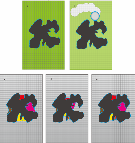
Schematic diagram of cavity detection in CAVITY. a) Protein (black-colored) in grid box (green-colored). b) Using the eraser ball to remove grid points outside protein. c) “Vacant” grid points after erasing. Four cavities were shown in different colors. d) Shrink each cavity until the depth reach the minimal depth. e) Recover cavities to obtain the final result.

Reference:

Yaxia Yuan, Jianfeng Pei, Luhua Lai. Binding Site Detection and Druggability Prediction of Protein Targets for Structure-Based Drug Design. Current Pharmaceutical Design, 2013,19 (12), 2326-2333(8). Link.

Yaxia Yuan, Jianfeng Pei, Luhua Lai. LigBuilder 2: A Practical de Novo Drug Design Approach. J. Chem. Inf. Model., 2011, 51 (5), 1083-1091. Link.

CavPharmer

Receptor-based Pharmacophore Modeling.

Pharmacophores derived from three-dimensional structures of specific protein binding sites can provide useful information for analyzing protein-ligand interactions, which therefore will help guide computational drug design. Pharmacophore features and their spatial arrangements are usually used to describe a pharmacophore model. CavPharmer uses a receptor-based pharmacophore modeling program Pocket2 to automatically extract pharmacophore features within cavities. CavPharmer output 7 feature types as hydrophobic center, hydrogen bond donor, hydrogen bond acceptor, positive-charged center, negative-charged center, aromatic center and exclude volume, to make a pharmacophore model from a protein structure integral. Key features in the pharmacophore model are automatically reduced to a reasonable number. We used data from DUD database to test the efficiency of CavPharmer and a ligand-based pharmacophore modeling software LigandScout V2.02. Results show that for receptor-based pharmacophore modeling, CavPharmer outperforms LigandScout. (average AUC value 0.69 versus 0.63 in 38 cases) Overall, CavPharmer is an accurate pharmacophore modeling software and can be applied widely into different drug design cases.

CavPharmer can also identify hot spots in protein-protein interface using only an apo protein structure. Given similarities and differences between the essence of pharmacophore and hot spots for guiding design of chemical compounds, not only energetic but also spatial properties of protein-protein interface are used in CavPharmer for dealing with protein-protein interface.

CavPharmer has been applied to many studies and well reproduced previously published pharmacophore models in these cases. One notable feature of CavPharmer is that it can tolerate minor conformational changes on the protein side upon binding of different ligands to give a consistent pharmacophore model. For different proteins accommodating the same ligand, CavPharmer gives similar pharmacophore models, which opens the possibility to classify proteins with their binding features. The Pharmacophore models used in Pharmmapper 2017 server (http://lilab.ecust.edu.cn/pharmmapper) were generated using CavPharmer method.

Reference:

J Chen, LH Lai, Pocket v.2: Further Developments on Receptor-Based Pharmacophore Modeling, J. Chem. Inf. Model., 2006,46(6),2684-2691.Link.

Corrsite

Potential allosteric ligand binding site prediction

The concept of allostery was proposed in early 1960’s, which mainly refers to the fact that the function of an orthosteric site can be affected by a ligand bound to an allosteric site that is topographically different from the orthosteric site. Allostery regulates numerous biological processes, and the dysregulation of allosteric communication networks between allosteric and orthosteric sites can lead to various human diseases. Compared to drugs targeting orthosteric sites, drugs targeting allosteric sites have higher specificity, fewer side effects and a variety of regulatory types. However, discovery of allosteric drugs remains challenging, as it is still difficult to accurately predict allosteric sites and to elucidate the mechanism of allosteric regulation.

CorrSite2.0 is a new method for predicting allosteric sites, which ranks the potential ligand binding sites based on calculated motion correlations using the slow and fast modes. The program first uses CAVITY to find all the potential ligand binding pockets on the surface of the protein. Then it selects an orthosteric pocket from the pockets found by CAVITY or imports an orthosteric pocket from the outside, such as an orthosteric pocket defined by the residues within 4Å around the orthosteric ligand. Next, the program calculates the correlations and Z-scores between the orthosteric pocket and the remaining pockets excluding the orthosteric sites in the top 10 fast modes and the top 3 slow modes, respectively. Finally, it takes the maximum of the two Z-Scores corresponding to each pocket, and ranks these pockets according to Z-Score. Pockets with Z-Score greater than 0.5 are predicted as potential allosteric sites.

Compared with CorrSite1.0, CorrSite2.0 has a great improvement in the applicability and prediction accuracy. First of all, CorrSite1.0 can only use one chain for prediction, while CorrSite2.0 can not only use one chain for prediction, but also can use multiple chains for prediction. Secondly, the prediction accuracy of CorrSite2.0 has been greatly improved. In the CorrSite2.0 dataset I containing 36 allosteric sites, CorrSite2.0 and CorrSite1.0 could successfully predict 35 and 25 of them, respectively. In the independent test set CorrSite2.0 dataset II, CorrSite2.0 and CorrSite1.0 could successfully predict 18 and 12 of 20 allosteric sites, respectively.

Reference:

Juan Xie, Shiwei Wang, Youjun Xu, Minghua Deng, Luhua Lai. Uncovering the dominant motion modes of allosteric regulation improves allosteric site prediction. J. Chem. Inf. Model. Link.

Xiaomin Ma, Hu Meng, Luhua Lai. Motions of Allosteric and Orthosteric Ligand-Binding Sites in Proteins are Highly Correlated. J. Chem. Inf. Model., 2016, 56 (9), 1725-1733. Link.

Yaxia Yuan, Jianfeng Pei, Luhua Lai. Binding site detection and druggability prediction of protein targets for structure-based drug design. Current pharmaceutical design 19.12 (2013): 2326-2333. Link.

CovCys

Detecting druggable cysteine residues for covalent ligand design

CovCys is developed based on a comprehensive statistical analysis on covalent modified cysteine residues in protein structures. Compared to unmodified cysteine residues in the same protein structure, the covalently modified cysteine residues showed lower pKa values and higher solvent exposure. Such cysteine residues are usually located within or near one of many pockets detected by a cavity-detection program.

The current application of computational procedure for targetable cysteine prediction only requires the protein three-dimensional structure coordinates. A number of descriptors will be calculated based on the structure, including surface pockets detected by using CAVITY, pKa by using PROPKA, SASA by using Pops as well as adjacent amino acid compositions calculated based on ProODY package. The generated descriptors will be used as the input for a pre-trained SVM model to predict whether a cysteine residues are suitable for druglike covalent ligand design. The external validation data set (positive/negative: 1377/5185) from Cysteinome database was tested by CovCys with prediction accuracy of 0.73.

Currently, if a cysteine is not within a pocket, it will not be used to make prediction. Such cysteine maybe an active cysteine, but it is hard to design a proper ligand without a binding cavity. Meanwhile, if the pKa calculation failed, it is also not considered. Such cysteine could be in a form of di-sulfur bond or inside the protein.

To use CovCys, please upload your protein structure information in the computing page. The output results contains all cysteine residues within the protein and their probability to be a druggable covalent reside. The pKa, percentage of exposure and the average pKd value of the associated pocket are also reported.

Reference:

Zhang W, Pei J, Lai L. Statistical Analysis and Prediction of Covalent Ligand Targeted Cysteine Residues. J Chem Inf Model. 2017,57, 1453-1460. Link.

Yuan Y., Pei J., Lai L. Binding Site Detection and Druggability Prediction of Protein Targets for Structure-Based Drug Design. Current Pharmaceutical Design, 2013,19, 2326-2333. Link.

Bas, D. C.; Rogers, D. M.; Jensen, J. H. Very Fast Prediction and Rationalization of pKa Values for Protein–Ligand Complexes Proteins: Struct., Funct., Genet. 2008, 73, 765–783. Link.

Marino, S. M. Protein Flexibility and Cysteine Reactivity: Influence of Mobility on the H-Bond Network and Effects on Pka Prediction Protein J. 2014, 33, 323– 336. Link.

Cavallo, L.; Kleinjung, J.; Fraternali, F. Pops: A Fast Algorithm for Solvent Accessible Surface Areas at Atomic and Residue Level Nucleic Acids Res. 2003, 31, 3364–3366. Link.

Bakan, A.; Meireles, L. M.; Bahar, I. ProDy: Protein Dynamics Inferred from Theory and Experiments Bioinformatics 2011, 27, 1575–1577. Link.